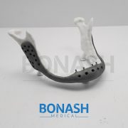

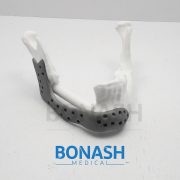
In this study we assessed the design criteria for the creation of a patient specific, whole mandible implant based on a patient’s medical imaging data and 3D printing. We tailor this procedure to a patient who will undergo a mandibulectomy due to cancer infiltration of the jaw. The patient CT scan data was used to generate a 3D representation of the patient’s skull, before the corrupted mandible was extracted. We examined two approaches based on classical symmetry matching and digital reconstruction of the defect to form the final model for printing. The final designs were then 3D printed and assessed for efficacy against a patient specific representative model of the skull and maxilla, where the final optimised design was found to provide an excellent fit. Ultimately, this technique provides a framework for the design and optimisation of a patient specific whole mandible implant.
Please note:
This abstract was published on Bonash Medical’s website since its content was related to the company’s products. There is no relation between Bonash Medical and the authors. To have full access to the article, please refer to relevant reference.

Custom implants for the reconstruction of craniofacial defects have gained importance due to better performance over their generic counterparts. This is due to the precise adaptation to the region of implantation, reduced surgical times and better cosmesis. Application of 3D modeling in craniofacial surgery is changing the way surgeons are planning surgeries and graphic designers are designing custom implants. Advances in manufacturing processes and ushering of additive manufacturing for direct production of implants has eliminated the constraints of shape, size and internal structure and mechanical properties making it possible for the fabrication of implants that conform to the physical and mechanical requirements of the region of implantation. This article will review recent trends in 3D modeling and custom implants in craniofacial reconstruction.
Please note:
This abstract was published on Bonash Medical’s website since its content was related to the company’s products. There is no relation between Bonash Medical and the authors. To have full access to the article, please refer to relevant reference.
Objective: To investigate the clinical application of the free fibula flap and the individualized titanium mesh, through virtual planning and guiding template to assist the reconstruction of maxilla and orbital floor defects.
Methods: Between 2015 and 2016, a total of six adult patients with maxillary and orbital floor defects were enrolled in the study. Preoperative virtual planning, including virtual maxillary resection and fibular reconstruction, was performed in all cases according to three-dimensional (3-D) radiographic and clinical findings. A printed 3-D resin model for pre-bent templates to guide the harvesting and positioning of the fibular flap during the surgery. Then an individualized titanium mesh is used to support the orbital floor and restore the maxillary contour. The results were confirmed by postoperative CT scans and clinical follow-up.
Results: Preoperative virtual planning and pre-bent template can be used to guide the harvesting and positioning of fibular flap and the forming and positioning of the individualized titanium mesh with satisfactory results. All flaps were survived and symmetric facial contours were achieved with normal lower jaw movements and proper vertical distance for dental implants in all patients.
Conclusion: Computer-aided techniques such as virtual planning, 3D printed model and pre-bending guide template can be used for the harvesting and positioning free fibula flap, the forming personalized titanium mesh, and ultimately the improving clinical efficacy of maxillary and orbital floor reconstruction.
Please note:
This abstract was published on Bonash Medical’s website since its content was related to the company’s products. There is no relation between Bonash Medical and the authors. To have full access to the article, please refer to relevant reference.
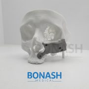
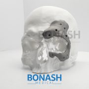
Purpose: To describe a novel technique using custom prostheses to repair fractured mandibular reconstruction plates spanning discontinuity defects.
Materials and Methods: This is a retrospective case series reviewing the design, rapid manufacture, and use of a novel method to repair fractured plates. Three patients who could not undergo autogenous bone grafting procedures or replacement of the entire plate for medical or socioeconomic factors were treated by this method.
Results: Three patients with fractured reconstruction plates were treated with a custom prosthesis engaging the reconstruction plate. Continuity and function were restored with a minimally invasive operation and short hospital stay. The custom prosthesis remained in place with stable occlusion in all 3 patients at a minimum of 9 months’ follow-up.
Conclusions: A technique using a custom prosthesis to quickly and less invasively restore continuity and function of the mandible after fracture of a reconstruction plate.
Please note:
This abstract was published on Bonash Medical’s website since its content was related to the company’s products. There is no relation between Bonash Medical and the authors. To have full access to the article, please refer to relevant reference.
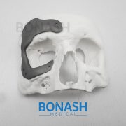
Customised implants manufacture has always presented difficulties which result in high cost and complex fabrication, mainly due to patients’ anatomical differences. The solution has been to produce prostheses with different sizes and use the one that best suits each patient. Additive manufacturing (AM) as a technology from engineering has been providing several advancements in the medical field, particularly as far as fabrication of implants is concerned in craniomaxillofacial surgery. The use of additive manufacturing in medicine has added, in an era of development of so many new technologies, the possibility of performing the surgical planning and simulation by using a threedimensional (3D) physical model, very faithful to the patient’s anatomy.
AM is a technology that enables the production of models and implants directly from the 3D virtual model (obtained by a Computer-Aided Design (CAD) system, computed tomography or magnetic resonance) facilitating surgical procedures and reducing risks. Furthermore, additive manufacturing has been used to produce implants especially designed for a particular patient, with sizes, shapes and mechanical properties optimised, for areas of medicine such as craniomaxillofacial surgery. This work presents how AM technologies were applied to design and fabricate a biomodel and customised implant for the surgical reconstruction of a large cranial defect. A series of computed tomography data was obtained and software was used to extract the cranial geometry. The protocol presented was used for creation of an anatomic biomodel of the bone defect for the surgical planning and, finally, the design and manufacture of the patient-specific implant.
Please note:
This abstract was published on Bonash Medical’s website since its content was related to the company’s products. There is no relation between Bonash Medical and the authors. To have full access to the article, please refer to relevant reference.
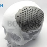
A diverse range of techniques is available for reconstruction of full-thickness calvarial defects and the optimum substrate for cranioplasty remains unproven. During a 9-year period, 149 patients underwent insertion of 151 custom-made titanium cranioplasties using the same technique. Data relating to patient demographics, indication for cranioplasty, and site and size of the defect were collected from the clinical records. Patients were followed up in all cases for a mean of 1 year 2 months (range 7 days to 8 years 8 months). Early complications requiring intervention were experienced in 7% and included seroma, haematoma, and continued bleeding necessitating implant removal in one patient.
One death occurred at 3 days’ post-operation due to haemorrhagic stroke. Late self-limiting complications such as seroma were experienced in 19% of patients, however complete failure requiring implant removal was seen in only 4% of cases. Infection was the cause of failure in all cases. A comprehensive literature review was carried out and data abstracted to compare reported failure rates in other techniques of full-thickness cranial reconstruction. This review shows that custom-made patient specific titanium cranioplasties compare very favorably to the other published techniques and remain a tried and tested option for reconstruction of all sizes of full-thickness calvarial defect.
Please note:
This abstract was published on Bonash Medical’s website since its content was related to the company’s products. There is no relation between Bonash Medical and the authors. To have full access to the article, please refer to relevant reference.