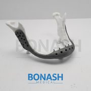

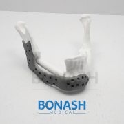
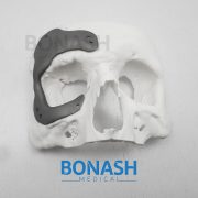
In this study we assessed the design criteria for the creation of a patient specific, whole mandible implant based on a patient’s medical imaging data and 3D printing. We tailor this procedure to a patient who will undergo a mandibulectomy due to cancer infiltration of the jaw. The patient CT scan data was used to generate a 3D representation of the patient’s skull, before the corrupted mandible was extracted. We examined two approaches based on classical symmetry matching and digital reconstruction of the defect to form the final model for printing. The final designs were then 3D printed and assessed for efficacy against a patient specific representative model of the skull and maxilla, where the final optimised design was found to provide an excellent fit. Ultimately, this technique provides a framework for the design and optimisation of a patient specific whole mandible implant.
Please note:
This abstract was published on Bonash Medical’s website since its content was related to the company’s products. There is no relation between Bonash Medical and the authors. To have full access to the article, please refer to relevant reference.
Objective: To investigate the clinical application of the free fibula flap and the individualized titanium mesh, through virtual planning and guiding template to assist the reconstruction of maxilla and orbital floor defects.
Methods: Between 2015 and 2016, a total of six adult patients with maxillary and orbital floor defects were enrolled in the study. Preoperative virtual planning, including virtual maxillary resection and fibular reconstruction, was performed in all cases according to three-dimensional (3-D) radiographic and clinical findings. A printed 3-D resin model for pre-bent templates to guide the harvesting and positioning of the fibular flap during the surgery. Then an individualized titanium mesh is used to support the orbital floor and restore the maxillary contour. The results were confirmed by postoperative CT scans and clinical follow-up.
Results: Preoperative virtual planning and pre-bent template can be used to guide the harvesting and positioning of fibular flap and the forming and positioning of the individualized titanium mesh with satisfactory results. All flaps were survived and symmetric facial contours were achieved with normal lower jaw movements and proper vertical distance for dental implants in all patients.
Conclusion: Computer-aided techniques such as virtual planning, 3D printed model and pre-bending guide template can be used for the harvesting and positioning free fibula flap, the forming personalized titanium mesh, and ultimately the improving clinical efficacy of maxillary and orbital floor reconstruction.
Please note:
This abstract was published on Bonash Medical’s website since its content was related to the company’s products. There is no relation between Bonash Medical and the authors. To have full access to the article, please refer to relevant reference.
Purpose: To describe a novel technique using custom prostheses to repair fractured mandibular reconstruction plates spanning discontinuity defects.
Materials and Methods: This is a retrospective case series reviewing the design, rapid manufacture, and use of a novel method to repair fractured plates. Three patients who could not undergo autogenous bone grafting procedures or replacement of the entire plate for medical or socioeconomic factors were treated by this method.
Results: Three patients with fractured reconstruction plates were treated with a custom prosthesis engaging the reconstruction plate. Continuity and function were restored with a minimally invasive operation and short hospital stay. The custom prosthesis remained in place with stable occlusion in all 3 patients at a minimum of 9 months’ follow-up.
Conclusions: A technique using a custom prosthesis to quickly and less invasively restore continuity and function of the mandible after fracture of a reconstruction plate.
Please note:
This abstract was published on Bonash Medical’s website since its content was related to the company’s products. There is no relation between Bonash Medical and the authors. To have full access to the article, please refer to relevant reference.
Customised implants manufacture has always presented difficulties which result in high cost and complex fabrication, mainly due to patients’ anatomical differences. The solution has been to produce prostheses with different sizes and use the one that best suits each patient. Additive manufacturing (AM) as a technology from engineering has been providing several advancements in the medical field, particularly as far as fabrication of implants is concerned in craniomaxillofacial surgery. The use of additive manufacturing in medicine has added, in an era of development of so many new technologies, the possibility of performing the surgical planning and simulation by using a threedimensional (3D) physical model, very faithful to the patient’s anatomy.
AM is a technology that enables the production of models and implants directly from the 3D virtual model (obtained by a Computer-Aided Design (CAD) system, computed tomography or magnetic resonance) facilitating surgical procedures and reducing risks. Furthermore, additive manufacturing has been used to produce implants especially designed for a particular patient, with sizes, shapes and mechanical properties optimised, for areas of medicine such as craniomaxillofacial surgery. This work presents how AM technologies were applied to design and fabricate a biomodel and customised implant for the surgical reconstruction of a large cranial defect. A series of computed tomography data was obtained and software was used to extract the cranial geometry. The protocol presented was used for creation of an anatomic biomodel of the bone defect for the surgical planning and, finally, the design and manufacture of the patient-specific implant.
Please note:
This abstract was published on Bonash Medical’s website since its content was related to the company’s products. There is no relation between Bonash Medical and the authors. To have full access to the article, please refer to relevant reference.
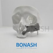
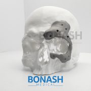
Reconstruction of the craniofacial complex is challenging because of the unique anatomy, the presence of vital structures and the diversity of defects. In craniofacial reconstruction, restoration of appearance and function is the primary goal. Autografts are the gold standard treatment, but they have several disadvantages, which has led to research into alloplastic materials. The development of CAD/CAM systems allows for precise preoperative planning and design of patient specific implants. In this process, two dimensional DICOM files were converted into 3-dimensional stereolithography files (STL) and the custom made titanium implant was designed using 3-dimensional software.
The skull and custom made titanium implant were printed as an STL model in resin for compatibility. The titanium implant was then printed using a laser sintering 3-dimensional printer.
We present the case of a patient who had his facial bones reconstructed because of a large deficiency in the ramus, body, and angle of his right mandible caused by an ameloblastoma.
Please note:
This abstract was published on Bonash Medical’s website since its content was related to the company’s products. There is no relation between Bonash Medical and the authors. To have full access to the article, please refer to relevant reference.
Abstract: The authors present the clinical results of their method of customized reconstruction of orbital wall defects using titanium mesh or sheet. High resolution computed tomography (CT) data are imported and processed to create a threedimensional (3D) image which is used to reconstruct the orbital defect. Mirror imaging of the air in the contralateral maxillary sinus is used to overcome artefact defects in the floor. A stereolithographic model is constructed, from which titanium mesh or sheet is shaped and sized to the required contours for implantation. Twentytwo patients were treated using this technique from 2003 to 2008. Postoperatively 10 patients reported early resolution of their diplopia. Six patients noticed significant improvement of their symptoms with mild residual diplopia in one direction only and at the extremes of gaze at final review. One patient required ocular muscle surgery. Enophthalmos resolved in eight of the nine cases. No patients developed enophthalmos or diplopia as a postoperative complication. The use of titanium mesh for orbital floor reconstruction has been shown to be safe and effective. Customized titanium implants accurately reproduce orbital contours thus restoring orbital volume. This reduces operative time and improves the functional and aesthetic outcomes of post-traumatic orbital reconstruction.
Please note:
This abstract was published on Bonash Medical’s website since its content was related to the company’s products. There is no relation between Bonash Medical and the authors. To have full access to the article, please refer to relevant reference.
The progress made in recent years in the field of head and neck bone reconstruction is directly related to technological advancements made in computeraided design and manufacturing (CAD/CAM) and three-dimensional printing in particular. Today these technologies are mainly used in mandibular reconstruction to manufacture aids for harvesting and shaping bone flaps. However problems remain when addressing patients with a contraindication to microsurgery who need extensive bone reconstruction. For these patients who cannot benefit from vascularized bone grafts, surgeons have to find alternative solutions aimed at maintaining best function and aesthetics. The goal of this article is to present an original method for mandibular body replacement with custom made porous titanium prostheses in patients ineligible for a bone free flap. This solution has been used for two patients with an intraoral approach, resulting in no visible scars, with simple postoperative care of a short duration. This innovative solution represents an additional option for the treatment of complex mandibular reconstructions.
Please note:
This abstract was published on Bonash Medical’s website since its content was related to the company’s products. There is no relation between Bonash Medical and the authors. To have full access to the article, please refer to relevant reference.