Custom implants in craniofacial surgery

3D modeling, custom implants and its future perspectives in craniofacial surgery

Dr. Jayanthi Parthasarathy, Dr, Plano

Annals of Maxillofacial Surgery

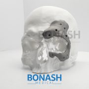
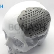
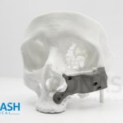
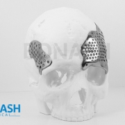
Custom implants for the reconstruction of craniofacial defects have gained importance due to better performance over their generic counterparts. This is due to the precise adaptation to the region of implantation, reduced surgical times and better cosmesis. Application of 3D modeling in craniofacial surgery is changing the way surgeons are planning surgeries and graphic designers are designing custom implants. Advances in manufacturing processes and ushering of additive manufacturing for direct production of implants has eliminated the constraints of shape, size and internal structure and mechanical properties making it possible for the fabrication of implants that conform to the physical and mechanical requirements of the region of implantation. This article will review recent trends in 3D modeling and custom implants in craniofacial reconstruction.

Please note:

This abstract was published on Bonash Medical’s website since its content was related to the company’s products. There is no relation between Bonash Medical and the authors. To have full access to the article, please refer to relevant reference.

Craniomaxillofacial surgery

Customized titanium implant fabricated in additive manufacturing for craniomaxillofacial surgery

André Luiz Jardini, Maria Aparecida Larosa, Cecília Amélia de Carvalho Zavaglia, Luis Fernando Bernardes, Carlos Salles Lambert, Paulo Kharmandayan, Davi Calderoni & Rubens Maciel Filho

Virtual and Physical Prototyping

Customised implants manufacture has always presented difficulties which result in high cost and complex fabrication, mainly due to patients’ anatomical differences. The solution has been to produce prostheses with different sizes and use the one that best suits each patient. Additive manufacturing (AM) as a technology from engineering has been providing several advancements in the medical field, particularly as far as fabrication of implants is concerned in craniomaxillofacial surgery. The use of additive manufacturing in medicine has added, in an era of development of so many new technologies, the possibility of performing the surgical planning and simulation by using a threedimensional (3D) physical model, very faithful to the patient’s anatomy.

AM is a technology that enables the production of models and implants directly from the 3D virtual model (obtained by a Computer-Aided Design (CAD) system, computed tomography or magnetic resonance) facilitating surgical procedures and reducing risks. Furthermore, additive manufacturing has been used to produce implants especially designed for a particular patient, with sizes, shapes and mechanical properties optimised, for areas of medicine such as craniomaxillofacial surgery. This work presents how AM technologies were applied to design and fabricate a biomodel and customised implant for the surgical reconstruction of a large cranial defect. A series of computed tomography data was obtained and software was used to extract the cranial geometry. The protocol presented was used for creation of an anatomic biomodel of the bone defect for the surgical planning and, finally, the design and manufacture of the patient-specific implant.

Please note:

This abstract was published on Bonash Medical’s website since its content was related to the company’s products. There is no relation between Bonash Medical and the authors. To have full access to the article, please refer to relevant reference.

Custom made cranioplasty

Custom made titanium cranioplasty early and late complications of 151 cranioplasties and review of the literature.

R. Williams, K. F. Fan, R. P. Bentley

Int. J. Oral Maxillofac. Surg.

A diverse range of techniques is available for reconstruction of full-thickness calvarial defects and the optimum substrate for cranioplasty remains unproven. During a 9-year period, 149 patients underwent insertion of 151 custom-made titanium cranioplasties using the same technique. Data relating to patient demographics, indication for cranioplasty, and site and size of the defect were collected from the clinical records. Patients were followed up in all cases for a mean of 1 year 2 months (range 7 days to 8 years 8 months). Early complications requiring intervention were experienced in 7% and included seroma, haematoma, and continued bleeding necessitating implant removal in one patient.

One death occurred at 3 days’ post-operation due to haemorrhagic stroke. Late self-limiting complications such as seroma were experienced in 19% of patients, however complete failure requiring implant removal was seen in only 4% of cases. Infection was the cause of failure in all cases. A comprehensive literature review was carried out and data abstracted to compare reported failure rates in other techniques of full-thickness cranial reconstruction. This review shows that custom-made patient specific titanium cranioplasties compare very favorably to the other published techniques and remain a tried and tested option for reconstruction of all sizes of full-thickness calvarial defect.

Please note:

This abstract was published on Bonash Medical’s website since its content was related to the company’s products. There is no relation between Bonash Medical and the authors. To have full access to the article, please refer to relevant reference.

Cranioplasty with Patient Specific Implants

Interval cranioplasty with patient specific implants and autogenous bone grafts – Success and cost analysis

Bernd Lethaus a, Monique Bloebaum a, David Koper a, Mariel Poort-ter Laak b, Peter Kessler

Journal of Cranio-Maxillo-Facial Surgery

Different options exist for the reconstruction of craniectomy defects following interval cranioplasty. The standard procedure is still based on the re-implantation of autogenous bone specimen which can be stored in the abdominal wall or be cryopreserved. Alternatively, patient-specific implants (PSIs) can be used. We conducted a retrospective study based on 50 consecutive patients with skull bone defects of 100 cm2 or more being operated on by the same team of surgeons. Thirty-three patients agreed to take part in the study. Seventeen patients who underwent reconstruction with PSIs (follow-up, 43 months [range, 3-93]) were compared with 16 control subjects who had autogenous bone grafts re-implanted (follow-up, 32 months [range, 5-92]). Criteria analyzed were the success and complication rates, operation time, duration of hospitalization and the treatment costs. Complication rate and the rate of reoperation were significantly lower, and the hospital stay was shorter in the PSI group. The treatment costs for reconstruction with autogenous bone were lower than skull bone reconstruction based on PSIs. Due to biological reasons some of the autogenous bone implants fail due to infection and resorption and the patients have to undergo another operation with implantation of a PSI in a secondary attempt. For those patients the highest overall treatment costs must be calculated. Conclusion: High success rates and reliability of PSIs may change the treatment strategy in patients undergoing interval cranioplasty. Please note: This abstract was published on Bonash Medical’s website since its content was related to the company’s products. There is no relation between Bonash Medical and the authors. To have full access to the article, please refer to relevant reference.

Cranial reconstruction

Cranial reconstruction: 3D biomodel and custom-built implant created using additive manufacturing.

André Luiz Jardinia,b, Maria Aparecida Larosaa,b*, Rubens Maciel Filhoa,b, Cecília Amélia de Carvalho Zavagliaa,c, Luis Fernando Bernardesa,b, Carlos Salles Lamberta,d, Davi Reis Calderonia,e, Paulo.

Journal of Cranio-Maxillo-Facial Surgery

Summary: Additive manufacturing (AM) technology from engineering has helped to achieve several advances in the medical field, particularly as far as fabrication of implants is concerned. The use of AM has made it possible to carry out surgical planning and simulation using a three-dimensional physical model which accurately represents the patient’s anatomy. AM technology enables the production of models and implants directly from a 3D virtual model, facilitating surgical procedures and reducing risks. Furthermore, AM has been used to produce implants designed for individual patients in areas of medicine such as craniomaxillofacial surgery, with optimal size, shape and mechanical properties. This work presents AM technologies which were applied to design and fabricate a biomodel and customized implant for the surgical reconstruction of a large cranial defect. A series of computed tomography data was obtained and software was used to extract the cranial geometry. The protocol presented was used to create an anatomic biomodel of the bone defect for surgical planning and, finally, the design and manufacture of the patient-specific implant.

Please note:

This abstract was published on Bonash Medical’s website since its content was related to the company’s products. There is no relation between Bonash Medical and the authors. To have full access to the article, please refer to relevant reference.

Patient Matched Implant for Cranioplasty

Patient Matched Implant for Cranioplasty

To have a patient matched implant for cranioplasty, the customizable nature of Patient specific implant lends itself to manufacture, ship, and autoclave at a competitive price point. Custom made implants benefit from many of the same advantages as Digital Surgery Planning (for example, CMF reconstruction) in that the advanced preparation can reduce operating room time, reduce risk, and help improve surgical outcomes.
Aesthetically the titanium mesh needs to be hand-bent to best fit the patient, but continual bending of titanium can contribute to micro-fractures that may compromise material strength. Work-hardening of titanium can lead to implant failure, but in the case of custom made implants, the surgeon could avoid the risk of work-hardening by selecting a 3d printed implant.

Cranial Custom Made Implant

Cranial Custom Made Implant

Custom made implant for the reconstruction of craniofacial defects have gained importance due to better performance over their generic counterparts. This is due to the precise adaptation to the region of implantation, reduced surgical times and better cosmetics of additively manufactured implants.

Advances in manufacturing processes for direct 3D printing of Patient specific implants has eliminated the constraints of shape, size and internal structure and mechanical properties making it possible for the fabrication of titanium implants that conform to the physical and mechanical requirements of the region of implantation.

In the vast majority of cases, precise symmetric reconstruction of maxillofacial defects remains an unsolved problem for craniofacial surgeons. Patient specific implants have contributed considerably to improvement in the accuracy and reliability of facial rehabilitation, rapidly becoming an irreplaceable part of the surgical armamentarium.

3d printed implants could be an advantageous and promising alternative to the use of other alloplastic materials. Moreover, custom made implants has the potential not only to achieve predictable correction for congenital or acquired deformities but also to serve a merely cosmetic purpose.

A titanium custom made chest wall implant could be a viable alternative for patients who had large chest wall tumors.