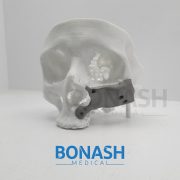
Reconstruction of maxilla
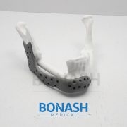
Reconstruction of maxillary and orbital floor defect with free fibula flap and whole individualized titanium mesh assisted by computer techniques.
Kun Fu, MDS, Yiming Liu, DDS, Ning Gao, MDS, Jinghua Cai, Phd, Wei He, DDS, Professor, Weiliu Qiu, DDS, Professor.
Journal of Oral and Maxillofacial Surgery
Objective: To investigate the clinical application of the free fibula flap and the individualized titanium mesh, through virtual planning and guiding template to assist the reconstruction of maxilla and orbital floor defects.
Methods: Between 2015 and 2016, a total of six adult patients with maxillary and orbital floor defects were enrolled in the study. Preoperative virtual planning, including virtual maxillary resection and fibular reconstruction, was performed in all cases according to three-dimensional (3-D) radiographic and clinical findings. A printed 3-D resin model for pre-bent templates to guide the harvesting and positioning of the fibular flap during the surgery. Then an individualized titanium mesh is used to support the orbital floor and restore the maxillary contour. The results were confirmed by postoperative CT scans and clinical follow-up.
Results: Preoperative virtual planning and pre-bent template can be used to guide the harvesting and positioning of fibular flap and the forming and positioning of the individualized titanium mesh with satisfactory results. All flaps were survived and symmetric facial contours were achieved with normal lower jaw movements and proper vertical distance for dental implants in all patients.
Conclusion: Computer-aided techniques such as virtual planning, 3D printed model and pre-bending guide template can be used for the harvesting and positioning free fibula flap, the forming personalized titanium mesh, and ultimately the improving clinical efficacy of maxillary and orbital floor reconstruction.
Please note:
This abstract was published on Bonash Medical’s website since its content was related to the company’s products. There is no relation between Bonash Medical and the authors. To have full access to the article, please refer to relevant reference.