Posts

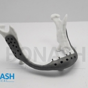
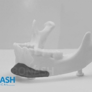
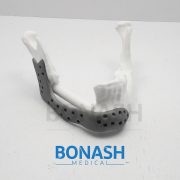
Custom made titanium implant

Reconstruction of complex mandibular defects using integrated dental custom-made titanium implants

A.Rachmiel, D.Shiloa, O.Blanca, O.Emodia,

British Journal of Oral and Maxillofacial Surgery.

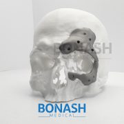
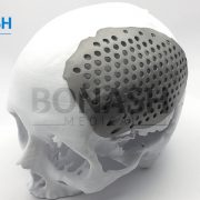
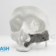
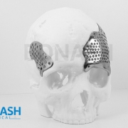
Reconstruction of the craniofacial complex is challenging because of the unique anatomy, the presence of vital structures and the diversity of defects. In craniofacial reconstruction, restoration of appearance and function is the primary goal. Autografts are the gold standard treatment, but they have several disadvantages, which has led to research into alloplastic materials. The development of CAD/CAM systems allows for precise preoperative planning and design of patient specific implants. In this process, two dimensional DICOM files were converted into 3-dimensional stereolithography files (STL) and the custom made titanium implant was designed using 3-dimensional software.

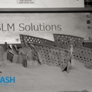
The skull and custom made titanium implant were printed as an STL model in resin for compatibility. The titanium implant was then printed using a laser sintering 3-dimensional printer.

We present the case of a patient who had his facial bones reconstructed because of a large deficiency in the ramus, body, and angle of his right mandible caused by an ameloblastoma.

Please note:

This abstract was published on Bonash Medical’s website since its content was related to the company’s products. There is no relation between Bonash Medical and the authors. To have full access to the article, please refer to relevant reference.

Orbital wall defects

Customized titanium reconstruction of post-traumatic orbital wall defects: a review of 22 cases

S. F. Mustafa, P. L. Evans, A. Bocca, D. W. Patton, A. W. Sugar, P. W. Baxter.

Int. J. Oral Maxillofac. Surg. 2011; 40: 1357–1362

Abstract: The authors present the clinical results of their method of customized reconstruction of orbital wall defects using titanium mesh or sheet. High resolution computed tomography (CT) data are imported and processed to create a threedimensional (3D) image which is used to reconstruct the orbital defect. Mirror imaging of the air in the contralateral maxillary sinus is used to overcome artefact defects in the floor. A stereolithographic model is constructed, from which titanium mesh or sheet is shaped and sized to the required contours for implantation. Twentytwo patients were treated using this technique from 2003 to 2008. Postoperatively 10 patients reported early resolution of their diplopia. Six patients noticed significant improvement of their symptoms with mild residual diplopia in one direction only and at the extremes of gaze at final review. One patient required ocular muscle surgery. Enophthalmos resolved in eight of the nine cases. No patients developed enophthalmos or diplopia as a postoperative complication. The use of titanium mesh for orbital floor reconstruction has been shown to be safe and effective. Customized titanium implants accurately reproduce orbital contours thus restoring orbital volume. This reduces operative time and improves the functional and aesthetic outcomes of post-traumatic orbital reconstruction.

Please note:

This abstract was published on Bonash Medical’s website since its content was related to the company’s products. There is no relation between Bonash Medical and the authors. To have full access to the article, please refer to relevant reference.

Cranial reconstruction

Cranial reconstruction: 3D biomodel and custom-built implant created using additive manufacturing.

André Luiz Jardinia,b, Maria Aparecida Larosaa,b*, Rubens Maciel Filhoa,b, Cecília Amélia de Carvalho Zavagliaa,c, Luis Fernando Bernardesa,b, Carlos Salles Lamberta,d, Davi Reis Calderonia,e, Paulo.

Journal of Cranio-Maxillo-Facial Surgery

Summary: Additive manufacturing (AM) technology from engineering has helped to achieve several advances in the medical field, particularly as far as fabrication of implants is concerned. The use of AM has made it possible to carry out surgical planning and simulation using a three-dimensional physical model which accurately represents the patient’s anatomy. AM technology enables the production of models and implants directly from a 3D virtual model, facilitating surgical procedures and reducing risks. Furthermore, AM has been used to produce implants designed for individual patients in areas of medicine such as craniomaxillofacial surgery, with optimal size, shape and mechanical properties. This work presents AM technologies which were applied to design and fabricate a biomodel and customized implant for the surgical reconstruction of a large cranial defect. A series of computed tomography data was obtained and software was used to extract the cranial geometry. The protocol presented was used to create an anatomic biomodel of the bone defect for surgical planning and, finally, the design and manufacture of the patient-specific implant.

Please note:

This abstract was published on Bonash Medical’s website since its content was related to the company’s products. There is no relation between Bonash Medical and the authors. To have full access to the article, please refer to relevant reference.

Custom made porous titanium prostheses

Use of a three-dimensional custom-made porous titanium prosthesis for mandibular body reconstruction

Q. Qassemyar, N. Assouly, S. Temam, F. Kolb

Int. J. Oral Maxillofac. Surg. 2017; 46: 1248–1251

doi: 10.1016/j.ijom.2017.06.001

The progress made in recent years in the field of head and neck bone reconstruction is directly related to technological advancements made in computeraided design and manufacturing (CAD/CAM) and three-dimensional printing in particular. Today these technologies are mainly used in mandibular reconstruction to manufacture aids for harvesting and shaping bone flaps. However problems remain when addressing patients with a contraindication to microsurgery who need extensive bone reconstruction. For these patients who cannot benefit from vascularized bone grafts, surgeons have to find alternative solutions aimed at maintaining best function and aesthetics. The goal of this article is to present an original method for mandibular body replacement with custom made porous titanium prostheses in patients ineligible for a bone free flap. This solution has been used for two patients with an intraoral approach, resulting in no visible scars, with simple postoperative care of a short duration. This innovative solution represents an additional option for the treatment of complex mandibular reconstructions.


Please note:

This abstract was published on Bonash Medical’s website since its content was related to the company’s products. There is no relation between Bonash Medical and the authors. To have full access to the article, please refer to relevant reference.

Patient Matched Implant for Cranioplasty

Patient Matched Implant for Cranioplasty

To have a patient matched implant for cranioplasty, the customizable nature of Patient specific implant lends itself to manufacture, ship, and autoclave at a competitive price point. Custom made implants benefit from many of the same advantages as Digital Surgery Planning (for example, CMF reconstruction) in that the advanced preparation can reduce operating room time, reduce risk, and help improve surgical outcomes.
Aesthetically the titanium mesh needs to be hand-bent to best fit the patient, but continual bending of titanium can contribute to micro-fractures that may compromise material strength. Work-hardening of titanium can lead to implant failure, but in the case of custom made implants, the surgeon could avoid the risk of work-hardening by selecting a 3d printed implant.

Cranial Custom Made Implant

Cranial Custom Made Implant

Custom made implant for the reconstruction of craniofacial defects have gained importance due to better performance over their generic counterparts. This is due to the precise adaptation to the region of implantation, reduced surgical times and better cosmetics of additively manufactured implants.

Advances in manufacturing processes for direct 3D printing of Patient specific implants has eliminated the constraints of shape, size and internal structure and mechanical properties making it possible for the fabrication of titanium implants that conform to the physical and mechanical requirements of the region of implantation.

In the vast majority of cases, precise symmetric reconstruction of maxillofacial defects remains an unsolved problem for craniofacial surgeons. Patient specific implants have contributed considerably to improvement in the accuracy and reliability of facial rehabilitation, rapidly becoming an irreplaceable part of the surgical armamentarium.

3d printed implants could be an advantageous and promising alternative to the use of other alloplastic materials. Moreover, custom made implants has the potential not only to achieve predictable correction for congenital or acquired deformities but also to serve a merely cosmetic purpose.

A titanium custom made chest wall implant could be a viable alternative for patients who had large chest wall tumors.

3D Printed Implants

Adaptation of bone implants with 3-D printing

One way to minimize the disparity between the material properties of the bone and 3D printed implants (patient specific implants) is match the mechanical properties and the stiffness. By inserting pores in the 3D printed titanium implants, the elastic modulus of the material can be reduced and bring closer to bone.
Using additive manufacturing techniques, 3d printed titanium implants with desired porosity can be predesigned which are also tailored for wall thickness, pore size, and bulk modulus.

For a patient-matched cranioplasty, the customizable nature of Patient specific implant lends itself to manufacture, ship, and autoclave at a competitive price point.  Custom made implants benefit from many of the same advantages as Digital Surgery Planning (for example, CMF reconstruction) in that the advanced preparation can reduce operating room time, reduce risk, and help improve surgical outcomes.

Porous Patient Specific Implants (PSI) are comparable to cortical bone and designed from patient scan data to create a customized, patient-specific solution. 3d printed implants are commonly used as an alternative to bone flaps to fill a defect or void in the cranial skeleton.

With extensive experience in computer-aided design and equipped with the latest 3D technologies, Bonash can supply you with a wide range of custom made implant.

Use of CAM/CAM in Implants Designing

Use of CAM/CAM in Implants Design

CAM/CAM systems have enabled us the ability to design and manufacture custom implants at an acceptable cost in a reasonable time. Additive manufacturing (3D printing) technologies lend themselves to manufacturing of complex 3d printed patient specific (custom made) implants without any barriers of design constraints.

Computer-aided manufacturing (CAM) is the use of software to control machine tools and related ones in the manufacturing of workpieces. This is not the only definition for CAM, but it is the most common CAM may also refer to the use of a computer to assist in all operations of a manufacturing plant, including planning, management, transportation and storage. Its primary purpose is to create a faster production process and components and tooling with more precise dimensions and material consistency, which in some cases, uses only the required amount of raw material (thus minimizing waste), while simultaneously reducing energy consumption. CAM is used in many schools alongside computer-aided design (CAD) to create objects.

Metal 3d printing uses bio-compatible implantable materials as pure titanium, Ti6Al4V and chrome cobalt and facilitate the direct production of custom made implants (patient specific implants) with engineered properties that match properties of the tissues at the region of implantation.

3D Printed Implant Manufacturing

3D Printed Implant Manufacturing

Using the CT scans, Bonash is able to make patient specific (custom made) 3d printed implant despite the substantial damage to patient’s skull. Researchers see 3D printing as beneficial in very cases as the implant fits more snugly due to customization and less time is spent in the operating room.

In the end, the patient looks better too with the incision scar hiding behind the hair line. They are also resistant in the case of an impact. The surgery to insert a typical 3D printed implant (custom made implant) took only about four hours, and was very successful.

The use of 3Dprinting has enabled us to work with patient specific model with a fully embedded implant. In the era of patient specific implant designs, using topology optimization method in order to find the structurally optimized solution, can eliminate the uncertainty of choosing heuristic shapes for critical surgeries like mid-face reconstruction.

3D Printing promises to produce complex biomedical devices according to computer design using patient specific anatomical data. Since its initial use as pre-surgical visualization models and tooling molds, 3D Printing has slowly evolved to create Patient specific (custom made) implants, scaffolds for tissue engineering and etc.

Additive Manufacturing in Medical

Additive Manufacturing in Medical

Additive Manufacturing technology enables the production of 3D printed implants directly from a 3D virtual model, facilitating surgical procedures and reducing risks. Furthermore, AM has been used to produce custom made implants designed for individual patients in areas of medicine such as craniomaxillofacial surgery, with optimal size, shape and mechanical properties.

AM medical models have found application for planning treatment for complex surgery procedures, training, surgical simulation, diagnosis, design and manufacturing of patient specific implants as well as medical tools. SLM techniques have been applied in the production of custom made implants that meet the physical characteristics of each patient.