Maxillary & mandibular defects

Surgical reconstruction of maxillary & mandibular defects using a printed titanium mesh

Xiao-Feng Shan, DDS, MD, Associate Professor, Hui-Min Chen, DDS, MD, Attending Physician, Jie Liang, DDS, Resident, Jin-Wei Huang, DDS, Resident, Zhi-Gang Cai, DDS, DMD, Professor

Journal of Oral and Maxillofacial Surgery.

Purpose: Reconstruction of maxillary & mandibular defects with a printed titanium mesh using computer-assisted surgery (CAS) for the achievement of structural, esthetic, and functional goals. Methods: The investigators designed and implemented this prospective study comprising patients with maxillary or mandibular flaws who underwent reconstruction with a printed titanium mesh using CAS. After surgery, the preoperative design and postoperative outcome were evaluated using Geomagic Studio software. Results: The sample comprised two patients with maxillary defects and two with mandibular defects. A satisfactory contour was achieved in all patients. The rate of concordance between the preoperative design and postoperative outcome was more than 81 and 94% within 3 mm for the mandibular and maxillary reconstructions, respectively. Conclusion: The results of this study suggest that complicated maxillary and mandibular flaws can be satisfactorily reconstructed with customized printed titanium meshes using CAS. Please note: This abstract was published on Bonash Medical’s website since its content was related to the company’s products. There is no relation between Bonash Medical and the authors. To have full access to the article, please refer to relevant reference.

Patient specific orbital implant

Patient specific implants (PSI) in reconstruction of orbital floor and wall fractures

Thomas Gander a, *, Harald Essig a, Philipp Metzler a, Daniel Lindhorst a, Leander Dubois b,

Journal of Cranio-Maxillo-Facial Surgery

Fractures of the orbital wall and floor can be challenging due to the demanding three dimensional anatomy and limited intraoperative overview. Misfitting implants and inaccurate surgical technique may lead to visual disturbance and unaesthetic results. A new approach using individually manufactured titanium implants for daily routine is presented in the current paper.  Preoperative CT-scan data were processed to generate a 3D-reconstruction of the affected orbit using the mirrored non-affected orbit as template and the extent of the patient specific implant (PSI) was outlined and three landmarks were positioned on the planned implant in order to allow easy control of the implant’s position by intraoperative navigation. Superimposition allows the comparison of the postoperative result with the preoperative planning. Neither reoperation was indicated due to malposition of the implant and the ocular bulb nor visual impairments could be assessed. PSI allows precise reconstruction of orbital fractures by using a complete digital workflow and should be considered superior to manually bent titanium mesh implants. Please note: This abstract was published on Bonash Medical’s website since its content was related to the company’s products. There is no relation between Bonash Medical and the authors. To have full access to the article, please refer to relevant reference.

Cranioplasty with Patient Specific Implants

Interval cranioplasty with patient specific implants and autogenous bone grafts – Success and cost analysis

Bernd Lethaus a, Monique Bloebaum a, David Koper a, Mariel Poort-ter Laak b, Peter Kessler

Journal of Cranio-Maxillo-Facial Surgery

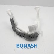
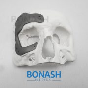
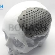
Different options exist for the reconstruction of craniectomy defects following interval cranioplasty. The standard procedure is still based on the re-implantation of autogenous bone specimen which can be stored in the abdominal wall or be cryopreserved. Alternatively, patient-specific implants (PSIs) can be used. We conducted a retrospective study based on 50 consecutive patients with skull bone defects of 100 cm2 or more being operated on by the same team of surgeons. Thirty-three patients agreed to take part in the study. Seventeen patients who underwent reconstruction with PSIs (follow-up, 43 months [range, 3-93]) were compared with 16 control subjects who had autogenous bone grafts re-implanted (follow-up, 32 months [range, 5-92]). Criteria analyzed were the success and complication rates, operation time, duration of hospitalization and the treatment costs. Complication rate and the rate of reoperation were significantly lower, and the hospital stay was shorter in the PSI group. The treatment costs for reconstruction with autogenous bone were lower than skull bone reconstruction based on PSIs. Due to biological reasons some of the autogenous bone implants fail due to infection and resorption and the patients have to undergo another operation with implantation of a PSI in a secondary attempt. For those patients the highest overall treatment costs must be calculated. Conclusion: High success rates and reliability of PSIs may change the treatment strategy in patients undergoing interval cranioplasty. Please note: This abstract was published on Bonash Medical’s website since its content was related to the company’s products. There is no relation between Bonash Medical and the authors. To have full access to the article, please refer to relevant reference.