Posts

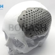
Custom made titanium implant

Reconstruction of complex mandibular defects using integrated dental custom-made titanium implants

A.Rachmiel, D.Shiloa, O.Blanca, O.Emodia,

British Journal of Oral and Maxillofacial Surgery.

Reconstruction of the craniofacial complex is challenging because of the unique anatomy, the presence of vital structures and the diversity of defects. In craniofacial reconstruction, restoration of appearance and function is the primary goal. Autografts are the gold standard treatment, but they have several disadvantages, which has led to research into alloplastic materials. The development of CAD/CAM systems allows for precise preoperative planning and design of patient specific implants. In this process, two dimensional DICOM files were converted into 3-dimensional stereolithography files (STL) and the custom made titanium implant was designed using 3-dimensional software.

The skull and custom made titanium implant were printed as an STL model in resin for compatibility. The titanium implant was then printed using a laser sintering 3-dimensional printer.

We present the case of a patient who had his facial bones reconstructed because of a large deficiency in the ramus, body, and angle of his right mandible caused by an ameloblastoma.

Please note:

This abstract was published on Bonash Medical’s website since its content was related to the company’s products. There is no relation between Bonash Medical and the authors. To have full access to the article, please refer to relevant reference.

Orbital wall defects

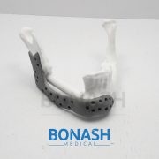
Customized titanium reconstruction of post-traumatic orbital wall defects: a review of 22 cases

S. F. Mustafa, P. L. Evans, A. Bocca, D. W. Patton, A. W. Sugar, P. W. Baxter.

Int. J. Oral Maxillofac. Surg. 2011; 40: 1357–1362

Abstract: The authors present the clinical results of their method of customized reconstruction of orbital wall defects using titanium mesh or sheet. High resolution computed tomography (CT) data are imported and processed to create a threedimensional (3D) image which is used to reconstruct the orbital defect. Mirror imaging of the air in the contralateral maxillary sinus is used to overcome artefact defects in the floor. A stereolithographic model is constructed, from which titanium mesh or sheet is shaped and sized to the required contours for implantation. Twentytwo patients were treated using this technique from 2003 to 2008. Postoperatively 10 patients reported early resolution of their diplopia. Six patients noticed significant improvement of their symptoms with mild residual diplopia in one direction only and at the extremes of gaze at final review. One patient required ocular muscle surgery. Enophthalmos resolved in eight of the nine cases. No patients developed enophthalmos or diplopia as a postoperative complication. The use of titanium mesh for orbital floor reconstruction has been shown to be safe and effective. Customized titanium implants accurately reproduce orbital contours thus restoring orbital volume. This reduces operative time and improves the functional and aesthetic outcomes of post-traumatic orbital reconstruction.

Please note:

This abstract was published on Bonash Medical’s website since its content was related to the company’s products. There is no relation between Bonash Medical and the authors. To have full access to the article, please refer to relevant reference.
Cranial reconstruction

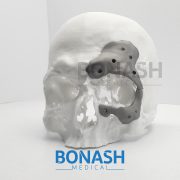
Cranial reconstruction: 3D biomodel and custom-built implant created using additive manufacturing.

André Luiz Jardinia,b, Maria Aparecida Larosaa,b*, Rubens Maciel Filhoa,b, Cecília Amélia de Carvalho Zavagliaa,c, Luis Fernando Bernardesa,b, Carlos Salles Lamberta,d, Davi Reis Calderonia,e, Paulo.

Journal of Cranio-Maxillo-Facial Surgery

Summary: Additive manufacturing (AM) technology from engineering has helped to achieve several advances in the medical field, particularly as far as fabrication of implants is concerned. The use of AM has made it possible to carry out surgical planning and simulation using a three-dimensional physical model which accurately represents the patient’s anatomy. AM technology enables the production of models and implants directly from a 3D virtual model, facilitating surgical procedures and reducing risks. Furthermore, AM has been used to produce implants designed for individual patients in areas of medicine such as craniomaxillofacial surgery, with optimal size, shape and mechanical properties. This work presents AM technologies which were applied to design and fabricate a biomodel and customized implant for the surgical reconstruction of a large cranial defect. A series of computed tomography data was obtained and software was used to extract the cranial geometry. The protocol presented was used to create an anatomic biomodel of the bone defect for surgical planning and, finally, the design and manufacture of the patient-specific implant.

Please note:

This abstract was published on Bonash Medical’s website since its content was related to the company’s products. There is no relation between Bonash Medical and the authors. To have full access to the article, please refer to relevant reference.